Procedures
Endoscopic surgery
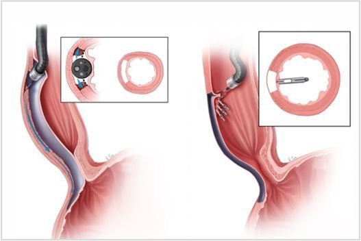
Endoscopic surgery is performed using a scope, a flexible tube with a camera and light at the tip. This allows doctor to see inside your colon and perform procedures without making major incisions, allowing for easier recovery time and less pain and discomfort. Endoscopic procedures are most often used for diagnosis.
Snaring is the most common surgical procedure that can be performed through any of the endoscopes. A snare is a wire that is like a lasso. The snare is looped over the tumor and tightened; then the wire is electrified to prevent bleeding as it cuts through.
Therapeutic ERCP
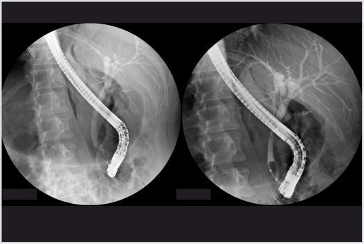
Endoscopic retrograde cholangiopancreatography, or ERCP, is a study of the ducts that drain the liver and pancreas. Ducts are drainage routes into the bowel. The ones that drain the liver and gallbladder are called bile or biliary ducts. The one that drains the pancreas is called the pancreatic duct. The bile and pancreatic ducts join together just before they drain into the upper bowel, about 3 inches from the stomach. The drainage opening is called the papilla. The papilla is surrounded by a circular muscle, called the sphincter of Oddi.
Diagnostic ERCP is when X-ray contrast dye is injected into the bile duct, the pancreatic duct, or both. This contrast dye is squirted through a small tube called a catheter that fits through the ERCP endoscope. X-rays are taken during ERCP to get pictures of these ducts. That is called diagnostic ERCP. However, most ERCPs are actually done for treatment and not just picture taking. When an ERCP is done to allow treatment, it is called therapeutic ERCP.
Endoscopic Ultrasound (EUS)
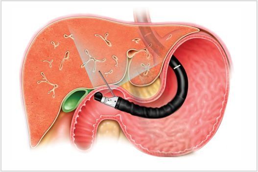
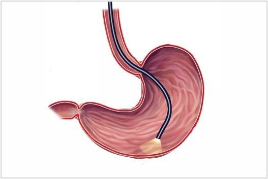
Endoscopic Ultrasound (EUS) is a minimally invasive procedure to assess gastrointestinal and Hepato-Pancreato- Billiary diseases. A special endoscope uses high-frequency sound waves to produce detailed images of the lining and walls of your digestive tract and chest, nearby organs such as the pancreas and liver, and lymph nodes.
When combined with a procedure called fine-needle aspiration (FNA) or FNB, EUS allows sampling (biopsy) fluid and tissue from abdomen (organ / lymph node) or chest for analysis. EUS with fine-needle aspiration can be a minimally invasive alternative to exploratory surgery.
Spiral Enteroscopy
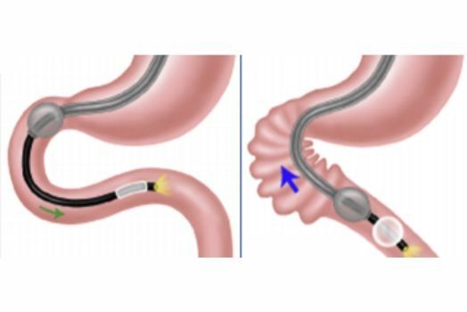
Spiral enteroscopy is a novel technique for small bowel exploration. The aim of this study is to compare double-balloon and spiral enteroscopy in patients with suspected small bowel lesions. The technique is safe and effective for the management and detection of small bowel pathology. Recent studies of spiral enteroscopy have demonstrated diagnostic yield, total time of procedure, and depth of insertion that compare favorably with double- and single-balloon enteroscopy. Spiral enteroscopy can be performed in postgastric surgery patients including Roux-en-Y patients requiring endoscopic retrograde cholangiopancreatography. Retrograde spiral enteroscopy can also be performed successfully. The strengths of spiral enteroscopy are rapid advancement in the small bowel and controlled, stable withdrawal that facilitates therapy. Future studies will be needed to compare competing technologies.
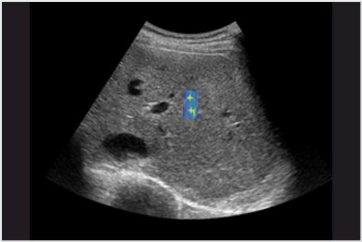
Liver Elastography / Fibro Scan
Liver elastography procedures help to determine the severity of hepatic fibrosis in patients with chronic liver disease. The Fibro Scan in particular can also help determine if a HCV patient does or does not have advanced fibrosis / cirrhosis, which can assist with treatment decisions regarding the need for oral antiviral therapy, liver cancer surveillance, etc. Obtaining serial liver elastography measurements once a year to see if the liver disease is improving or worsening over time.
Fibro Scan test results are always used in conjunction with other clinical data, laboratory test results, and liver imaging in managing individual patients. The Fibro Scan test is completely non-invasive, painless procedure that takes approximately 10 minutes to complete. Most patients with chronic liver disease can be assessed.
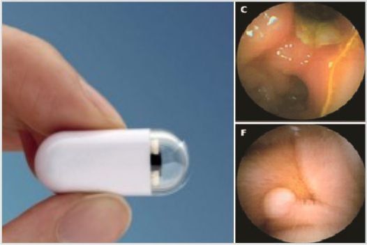
Capsule Endoscopy
Capsule endoscopy is a procedure used to record internal images of the gastrointestinal tract for use in medical diagnosis. A capsule endoscopy camera sits inside a vitamin-size capsule you swallow. As the capsule travels through your digestive tract, the camera takes thousands of pictures that are transmitted to a recorder you wear on a belt around your waist.
Capsule endoscopy helps doctors see inside your small intestine — an area that isn't easily reached with more-traditional endoscopy procedures. Traditional endoscopy involves passing a long, flexible tube equipped with a video camera down your throat or through your rectum. Newer developments are also able to take biopsies and release medication at specific locations of the entire gastrointestinal tract.
Endoscopic Management of Obesity
Endoscopic management of obesity can be grouped under the following 3 categories:
- primary management of obesity
- management of obesity-related disorders such as diabetes mellitus and infertility
- management of postsurgical complications
At present, endoscopic treatment is preferred over surgery for postsurgical complications, such as bleeding, dilation of strictures anastomosis, constriction of a dilated anastomosis, and occasionally fistula formation.
Currently there are three types of Endoscopic management of obesity used for the treatment of obesity:
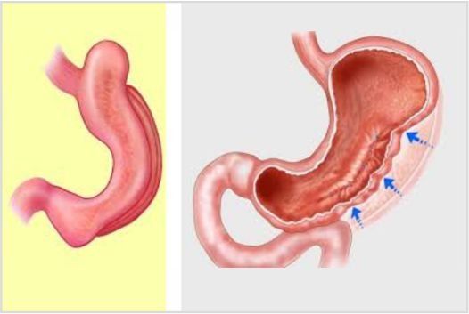
- Intragastric balloons: An intragastric balloon is a space-occupying device that is placed in the stomach. The mechanism of action of Intragastric balloons involves delaying gastric emptying, which leads to increased satiety.
- Plication and suturing
- Aspiration therapy
Endoscopy
A Endoscopy is an examination that allows us to look directly at the upper part of the gastrointestinal tract; the oesophagus (tube that food passes down to reach the stomach); the stomach and around the first bend of the small intestine (duodenum).
- In order to do this, a thin flexible tube called a gastroscope or endoscope, which has a light at one end, is used. It is passed through the mouth, down the oesophagus and into the stomach by a specially trained doctor. The tube is thinner than your little finger. It will not get in the way of your breathing at any time, as it passes down your oesophagus and not your windpipe.
- Sometimes biopsies (small tissue samples) are taken and sent for analysis. The biopsies taken are about the size of a match head and will not cause you any pain. You may feel a slight pull sensation.
Colonoscopy
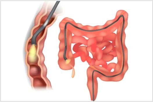
A colonoscopy is a routine test to examine the lining of your bowel, also called the large intestine or colon. This is done using an endoscope, which is a flexible tube, about the thickness of a (little) finger, which has a camera and light at one end. It is passed through the anus (back passage) and carefully moved around the large bowel by a specially trained doctor.
In case of abdominal pain , gastritis, ulcers, bleeding in digestive tract, bowel habit changes (diarrhea or chronic constipation), colon growth or polyps, chronic fatigue, blood in stools, narrow/thin stools colonoscopy is suggested.
Hydrogen Breath Test
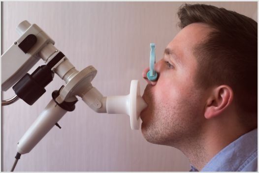
The hydrogen breath test is a test that uses the measurement of hydrogen in the breath to diagnose several conditions that cause gastrointestinal symptoms. Hydrogen breath testing is used in the diagnosis of three conditions:
- The first is a condition in which dietary sugars are not digested normally. The most common sugar that is poorly digested is lactose, the sugar in milk. Individuals who are unable to properly digest lactose are referred to as lactose intolerant. Testing also may be used to diagnose problems with the digestion of other sugars such as sucrose, fructose and sorbitol.
- The second condition for which hydrogen breath testing is used is for diagnosing bacterial overgrowth of the small bowel, a condition in which larger-than-normal numbers of colonic bacteria are present in the small intestine.
- The third condition for which hydrogen breath testing is used is for diagnosing rapid passage of food through the small intestine. All three of these conditions may cause abdominal pain, abdominal bloating and distention, flatulence (passing gas in large amounts), and diarrhea.
Sigmoidoscopy
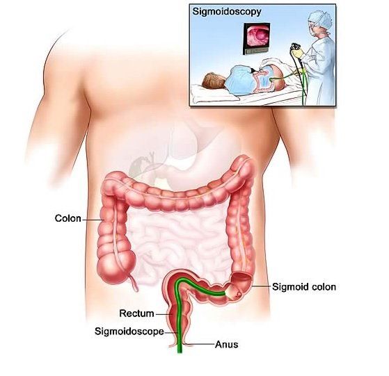
A sigmoidoscopy, also called a flexible sigmoidoscopy, is a procedure that lets doctor look inside your sigmoid colon (the rectum and lower part of the large intestine) by using a flexible tube with a light on it. It helps doctor check for:
- Ulcers
- Rectal bleeding
- Abnormal cells
- Chronic diarrhea
- Polyps
- Cancer
- Other intestinal problems
Sigmoidoscopy is one option for colon cancer screening, but there are other options that allow your doctor to view your whole colon, such as colonoscopy. Typically, pieces of tissue will be taken as samples to check for any abnormal cell changes.
Gastrointestinal Physiology Study
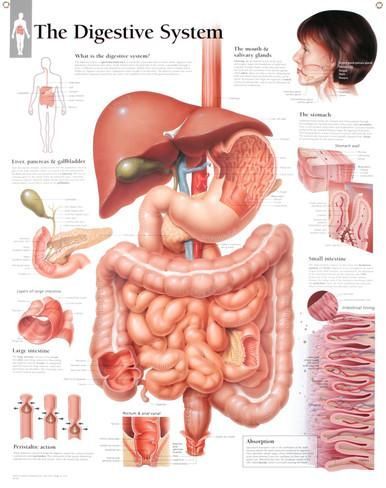
The gastrointestinal system consists of the gastrointestinal tract and the accessory exocrine glands. The gastrointestinal tract includes the mouth, the esophagus, the stomach, the small intestine, and the large intestine. The major accessory glands are the salivary glands, the liver, the gallbladder, and the pancreas. Gastrointestinal physiology concerns any part of the digestive tract from the oesophagus to the anus and also of the liver, biliary tract and the pancreas.
Liver Transplantation
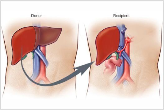
Liver transplantation surgically replaces a failing or diseased liver with one that is normal and healthy. At this time, transplantation is the only cure for liver insufficiency or liver failure because no device or machine reliably performs all of the functions of the liver. People who require liver transplants typically have one of the following conditions.
- Acute Liver Failure
- Chronic liver failure
There are many people with cirrhosis and decompensated liver disease but not all are appropriate candidates for liver transplantation. A patient must be able to survive the operation and the potential post-operative complications, reliably take the medications that prevent rejection and opportunistic infections, comply with frequent visits and laboratory tests, and not engage in activity that would injure the liver, such as drinking alcohol.


